My Baby Doesn' T Have Soft Tissues to Close Palate
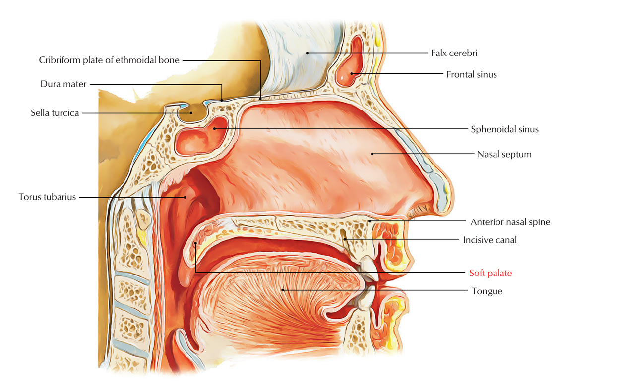
The soft palate is a movable muscular flap, which hangs down from the posterior margin of the hard palate into the pharyngeal cavity. It divides the nasopharynx from the oropharynx. The soft palate tin be easily distinguished from the hard palate at the front of the oral cavity since it does non comprise bone.
External Features
The soft palate presents the following external features:
- Anterior (oral) surface is concave as well as indicated by a median raphe.
- Posterior surface is convex and continuous with the floor of the nasal crenel.
- Superior edge is connected to the posterior edge of the hard palate.
- Inferior border is free and creates the inductive boundary of the pharyngeal isthmus.
- A conical, small, tongue like project hanging down from its middle is referred to as uvula.
On every side from the base of the uvula, 2 curved folds of mucous membrane stretch laterally and downwards:
- The inductive fold unites inferiorly with the side of the tongue (at the junction of oral and pharyngeal parts) and is chosen palatoglossal fold. The palatoglossal fold includes the palatoglossus muscle and creates the lateral boundary of the oropharyngeal isthmus.
- The posterior fold unites inferiorly with the lateral wall of the pharynx and is called palatopharyngeal fold. The palatopharyngeal fold includes palatopharyngeus muscle and creates the posterior boundary of the tonsillar fossa.
Structure
The soft palate is created from a fold of mucous membrane enclosing 5 pairs of muscles. The nasal surface of the soft palate is covered by pseudostratified ciliated columnar epithelium with the exception of posteriorly (the part that abuts on the Passavant's ridge of posterior pharyngeal wall) that is lined by non-keratinized stratified squamous epithelium. The oral surface of the soft palate is thicker and lined past non-keratinized stratified squamous epithelium.

Soft Palate
In the submucosa on both the surfaces are mucous glands, which are in loads around the uvula and on the oral attribute of the soft palate. The mucosa on the oral surface of the soft palate too includes some taste buds (specially in children) and lymphoid follicles.
Muscles
The soft palate is equanimous of the 5 pair of muscles, viz
- Tensor palati (tensor veli palatini).
- Levator palati (levator veli palatini).
- Palatoglossus.
- Palatopharyngeus.
- Musculus uvulae.
All the muscles of soft palate are extrinsic with the exception of musculus uvulae that are intrinsic.
Origin, Insertion and Activities of Musculus of The Soft Palate
| Muscle | Origin | Insertion | Actions |
|---|---|---|---|
| Tensor palati (sparse triangular musculus; | (a) Lateral aspect of the cartilaginous office of the auditory tube. (b) Adjoining part of the greater wing of the sphenoid including its spine | Muscle descends, converges to form a tendon, which hooks around thepterygoid hamulus and so expands to form the palatine aponeurosis for attachment to: Posterior edge of the hard palate Inferior surface of the difficult palate backside the palatine crest | (a) Tightens the soft palate. (b) Helps in opening the auditory tube |
| Levator palati (a cylindrical muscle lying deep to tensor palati) | (a) Medial aspect of the cartilaginous part of the auditory tube. (b) Adjoining part of the petrous temporal os (inferior surface of its noon anterior to carotid canal) | Musculus runs downwardly and medially and spreads out to be inserted on the upper surface of the palatine aponeurosis | (a) Elevates the soft palate to close the pharyngeal isthmus. (b) Helps in opening the auditory tube. |
| Muscle uvulae (a longitudinal musculus strip, one on either side of the median plane within the palatine aponeurosis) | (a) Posterior nasal spine. (b) Palatine aponeurosis | Mucous membrane of the uvula | Pulls the uvula forrad to its own side |
| Palatoglossus | Oral surface of the palatine aponeurosis | Descends into palatoglossal curvation, to be inserted into the side of the tongue at the junction of its oral and pharyngeal parts | (a) Pulls upwardly the root of the natural language. (b) Approximates the palatoglossal arches to close the oropharyngeal isthmus |
| Palatopharyngeus (consist of 2 fasciculi, which are separated by the levator palati) | (a) Inductive fasciculus: from posterior edge of the hard palate. (b) Posterior fasciculus: from palatine aponeurosis | Descends in the palatopharyngeal arch and inserted into the Median fibrous raphe of pharyngeal wall Posterior border of the lamina of thyroid cartilage | Raises the walls of throat and larynx during swallowing |
Functions of Soft Palate
- Separates the oropharynx from nasopharynx during swallowing so that nutrient will not become into the olfactory organ.
- Sequester the oral cavity from oropharynx during mastication and then that breathing isn't changed.
- Helps to change the attribute of voice, by altering the level of blockage of the pharyngeal isthmus.
- Shields the damage of nasal mucosa during sneezing, past suitably breaking upward and directing the gust of air via both nasal and oral cavities.
- Prevents the archway of sputum into olfactory organ during cough by directing it in the oral cavity.
Arterial Supply
The soft palate is supplied by the post-obit arteries:
- Lesser palatine branches of the maxillary avenue.
- Ascending palatine branch of the facial artery.
- Palatine branches of the ascending pharyngeal artery.
Venous Drainage
The venous blood from palate is drained into pharyngeal venous plexus and pterygoid venous plexus.
Lymphatic Drainage
The lymphatics from soft palate drain into retropharyngeal and upper deep cervical lymph nodes.
Nerve Supply
Motor Supply:
All the muscles of soft palate are supplied by the cranial root of accessory nerve via pharyngeal plexus with the exception of tensor palati, which is supplied by the nerve to medial pterygoid, a branch of the mandibular nerve.
Sensory Supply:
- General sensations from palate are carried by:
- Lesser palatine nerves to the maxillary partitioning of trigeminal nerve via pterygopalatine ganglion.
- Glossopharyngeal nerve.
Clinical Significance
Gag reflex
It's a protective reflex demarcated by the elevation of the palate and contraction of the pharyngeal muscles with related retching and gagging in response to stimulant of the mucous membrane of the oropharynx. It takes identify when the palate, tonsil, posterior role of the tongue, or posterior pharyngeal wall are contacted past unknown things like swab, spatula, etc. The afferent limb of the reflex is supplied by the glossopharyngeal nerve and efferent limb past the vagus nerve.
Paralysis of soft palate
The paralysis of the muscles of soft palate due to lesion of vagus nerve creates:
- Nasal regurgitation of liquids.
- Nasal twang in voice.
- Flattening of the palatal arch on the side of the lesion.
- Deviation of uvula, opposite to the side of the lesion.
Oropharyngeal Cancer
The junior aspect of the soft palate creates the upper part of the oropharynx and is the location of the where most of the soft palate cancers occur.
The superior surface of the soft palate i.e. portion of the nasopharynx, is rarely a primary site of neoplasm germination.
- The soft palate has a substantial amount of minor salivary glands.
- These glands act as the location of origin for non-squamous neoplasms of the soft palate.
- A soft palate tumor can extend anteriorly equally well as also encompass the hard palate.
- Perineural extend also announced superiorly within the pterygopalatine fossa alongside the greater equally well as lesser palatine divisions of the maxillary nerve.
Further perineural extension from the pterygopalatine fossa can cause extension:
- Via the inferior orbital fissure along divisions of the maxillary division of the trigeminal nerve inside the orbit.
- Via the foramen rotundum inside the key skull base.
- Via the nervus of the pterygoid culvert inside the facial nerve and temporal os.
Both the trigeminal and facial nerves palsies can appear due to this perineural spread.
[su_spoiler championship="References"]
- Gray'south Anatomy for Students Topic Soft Palate P. 267
- The Journal of family practice vol five P. 55
- The Cleft palate journal p. 134–61. 4524356
[/su_spoiler]
Source: https://www.earthslab.com/anatomy/soft-palate/
0 Response to "My Baby Doesn' T Have Soft Tissues to Close Palate"
Postar um comentário